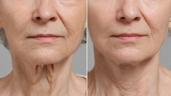
Injection of botulinum toxin type A (BTX-A) into the masseter muscle is becoming a common procedure performed by injectors. Hypertrophy of the masseter muscle results in the appearance of a square face and jaw, and this condition may be exacerbated by habits and conditions including teeth grinding, jaw clenching or bruxism. For patients who desire a slimmer-looking face or wish to correct this appearance of a square jaw, injectors can target the superficial region of the masseter muscle, which connects the mandible to the cheekbone and is involved in chewing and talking. In addition to cosmetic uses, BTX-A is an effective therapeutic treatment for conditions including temporomandibular disorder (TMD), masticatory myalgia and localized muscle hyperactivity.
While BTX-A treatment of the masseter muscle has clear clinical benefits, it has also been associated with long-term changes in mandibular bone morphology and reductions in bone quality, including reductions in skeletal muscle size as well as thinning and shrinkage of trabecular and cortical mandibular structures. These results are concerning, as they could potentially lead to irreversible unwanted changes in lower facial structure over time as well as impaired jaw function and a host of other clinical symptoms.
On the following pages, we will discuss potential mechanisms through which this treatment might produce these long-term effects on the surrounding bone and facial structure. With these considerations in mind, we discuss specific approaches that can be taken to maximize this treatment’s efficacy while limiting the risk of adverse consequences.
Bone Basics and Remodeling
Bone cells are connected by intercellular gap junctions, which allow them to rapidly transmit signals to each other and form an interconnected cellular network. Additionally, the bone matrix is highly vascularized. This direct connection to the systemic circulation allows bone cells to become sensitive to the climate of the body and respond to changes accordingly.
There are three major types of bone cells: osteoblasts, osteoclasts and osteocytes. Osteoblasts are responsible for bone formation while osteoclasts are responsible for bone resorption, both of which occur in remodeling. The bone is under a constant state of remodeling. This process is necessary to remove old or damaged bone tissue, maintain plasma calcium levels and adjust bone architecture. Consequently, there is constant communication between osteoblasts and osteoclasts to balance their opposing processes of bone formation and breakdown. The external signals and physiological changes induced by BTX-A treatment can lead to changes favoring activity of osteoclasts over that of osteoblasts; thereby resulting in net bone breakdown.
While we typically understand osteoclasts and osteoblasts as the major cells involved in bone homeostasis, osteocytes also play a significant role. These cells are terminally differentiated osteoblasts, typically found deep within mineralized bone. Osteocytes are involved in regulating processes of bone remodeling and mineral homeostasis as well as modifying their microenvironment. Osteocytes respond to a variety of signals, one of which is mechanical strain. This may shed light on how paralysis of the muscle, induced by BTX-A treatment, may lead to skeletal bone changes.
The impact and pressure to bone is translated into signals of mechanical strain, which can induce osteocytes to release factors that alter the activity of surrounding cells and initiate changes in bone remodeling processes.1 Therefore, masseter muscle paralysis may induce remodeling changes in mandibular bony structures that favor bone breakdown, giving rise to the bone thinning and different morphologies that have been seen.
The effect of muscle activity on bone density is multifactorial. Surrounding muscles are linked to the bone in four major ways: 1. anatomically through direct contact and connections; 2. mechanically through exertion of contractile forces on the bone; 3. metabolically through hormonal crosstalk and release of secretory factors; and 4. pleiotropically through shared phenotypic traits and genetic influences and pathways.2 This helps explain how alterations in muscle size, density and strength can be transmitted to related bone structures.
The body is constantly in the process of bone remodeling, but remodeling is also a response to the demands of mechanical loading. Mechanical stimulation of the muscles leads to the transduction of this mechanical loading to the bone, which is required to maintain bone density. This transduction of mechanical loading to the bone from the skeletal muscles is achieved in three ways: 1. production of tensile force by muscle contraction at its insertion site; 2. production of compressive force by the muscles that transduce loading across the joints; and 3. production of bending force from the long bone-associated muscles when lifting distally held objects.3
Differences in the magnitude, direction, rate and frequency of the mechanical load can all result in distinct changes in remodeling and bone development.2 Thus, mechanical load is a driver of healthy bone remodeling processes and is necessary to increase the strength and stability of the bone.
Continue Reading to learn more about BTX-A and bone loss in our Digital Magazine...
Kay Durairaj, MD, is the founder and medical director of Beauty by Dr. Kay, Pasadena, California; chair of the department of ENT at Huntington Memorial Hospital; and clinical professor at UCLA/Olive View Medical Center.
Tivoli Nguyen graduated from the University of Chicago with a degree in Biological Sciences and specialization in Endocrinology.
1. “Osteocyte.” Osteocyte - an Overview, ScienceDirect Topics, sciencedirect.com/topics/agricultural-and-biological-sciences/osteocyte.
2. Hart, N H, et al. “Mechanical Basis of Bone Strength: Influence of Bone Material, Bone Structure and Muscle Action.” Journal of Musculoskeletal & Neuronal Interactions, 1 Sept. 2017
3. Hong, Seok Woo, and Jeong-Hyun Kang. “Decreased Mandibular Cortical Bone Quality after Botulinum Toxin Injections in Masticatory Muscles in Female Adults.” Nature News, 27 Feb. 2020.