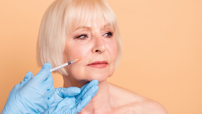
Dermal fillers, also called subdermal implants, are frequently used in aesthetic medicine. Along with an increase in the number of these procedures being performed, an increase in complications is evident. The most serious complication is vascular occlusion.
Treatment of vascular complications during their imminent presentation should be priority knowledge for all injectors. Every practitioner should have at hand an algorithm for the proper treatment of impending vascular complications with subdermal implants with the aim of ideally suppressing, or eventually minimizing, the potentially catastrophic impact of these complications, such as skin necrosis and blindness.
Vascular compromise manifests through three proposed mechanisms:
- Intravascular embolism
- Extravascular compression
- Vascular spasm
Intravascular embolism is the mechanism with best evidence. It is caused by inadvertent intravascular injection of filler material, leading to vascular occlusion and, eventually, necrosis. Some animal models have suggested that hyaluronic acid particles within a vessel may induce some degree of vascular spasm, reducing the necrosis area.
Keep in mind that some cases of vascular compromise may have a delayed presentation—a mechanism that is poorly understood but associated with filler migration to low diameter capillaries, hyaluronic acid’s hydrophilic nature, embolus induction of platelet aggregation or failure of collateral blood supply after an initial vascular injury.
Danger Zones
Several facial regions have been described as high-risk territories, but I believe that there are no “safe” facial areas. Every treatment must be performed with deep anatomical knowledge and attention to detail. That said, the areas at greatest risk of vascular compromise during and following the injection of dermal fillers include the glabellar region, nasolabial folds and the nose.
In the glabellar region we find the supratrochlear artery, supraorbital artery and cutaneous branches of the ophthalmic artery. This is one of the regions of the face where the internal carotid system connects with the external carotid system. Also, this area has poor collateral circulation.
The nasolabial fold is intimately related with the facial artery, and the risk of vascular compromise when injecting in this area is high.
The nose is another high-risk area, where the angular artery and dorsal nasal artery anastomose connect the internal and external carotid systems. The nasal tip and the alar triangle both rely on the lateral nasal artery solely, creating a poor collateral circulation zone.
Miguel A. Aristizábal, MD, is the co-founder of the ADEI - Aesthetics & Dermatology Institute in Bogota, Colombia.