A group of 17 international aesthetic experts with experience in injection of calcium hydroxylapatite (CaHA) and the management of CaHA‐related complications convened to develop a consensus on the optimal management of vascular compromise following intra‐arterial CaHA injection. Their consensus guidelines were published in the Journal of Cosmetic Dermatology (November 2020).
The paper covers high risk areas for CaHA injection, injection technique, how to recognize the symptoms of vascular occlusion leading to vision loss or tissue necrosis as well as detailed treatment protocols for the management of these events.
In addition to having a through knowledge of facial vascular anatomy, the consensus members noted that physicians should be aware that there are large variations between individuals in the branching patterns of many arteries. Patients who have undergone previous cosmetic procedures such as rhinoplasty may have unpredictable repositioning of blood vessels and a more tenuous blood supply in the operated area, and previous scars may also fix arteries in place, making them easier to penetrate with small sharp needles or even cannulae.
Risk Zones and Injection Technique
Areas of the face that have the highest risk of vascular occlusion include the nasal tip and alar. The glabella—the most common filler injection site triggering blindness—is a contraindication for CaHA, and the product should not be injected there.
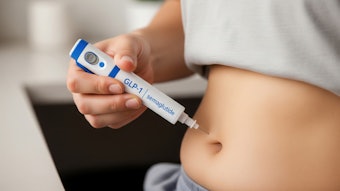
CaHA should be injected slowly with a low injection force using the smallest amount of product necessary. Injectors should avoid large boluses and limit injection volumes to 0.1 mL. If unexpected resistance is detected, blanching observed or the patient indicates pain, stop the injection immediately.
Recognizing and Responding to Vascular Occlusion
If an injection disrupts the blood supply to the retina—which can occur after dermal filler injection in almost any area of the face—the patient will experience a sudden onset of impairment/loss of vision accompanied by severe pain (ocular, facial, and/or headache). Because cerebral infarction can accompany retinal artery occlusion, signs and symptoms such as aphasia or even contralateral hemiparesis might also be present.
Once the retinal artery has been occluded, only a short window of opportunity of about 60 minutes exists before blindness is irreversible. The immediate goal is rapid reduction of intraocular pressure to allow the emboli to dislodge downstream and improve retinal perfusion. Treatment may include application of topical and systemic intraocular pressure lowering agents, isovolemic hemodilution, ocular massage, and aspirin anticoagulation, as well as carbon dioxide inhalation, oral corticosteroids and hyperbaric oxygen therapy. The patient must be transferred immediately to the nearest ophthalmologist with as much information as possible regarding the product, injection site(s) and the volume of injection.
In cases of vascular occlusion leading to tissue necrosis, the patient will experience disproportionate pain, though this may be masked due to the anesthetics mixed in certain fillers. Additional symptoms include pallor and blanching, which usually present themselves immediately, and/or mottled discoloration. Progressive symptoms include hemorrhagic changes (24‐48 hours), blisters and pustules (4‐6 days) and necrosis (5‐10 days). Finally, secondary infection may occur.
If you suspect the blood supply has been compromised, stop injecting immediately and, if possible, try to aspirate the product before withdrawing the needle. Apply warm compresses and massage the area to promote vasodilation and perfusion.
Inject hyaluronidase, which the panel notes should be used regardless of the filler type due to its edema‐reducing and anti‐inflammatory properties, directly into the affected site. There was strong consensus for high doses (600U of hyaluronidase for every 0.1 mL CaHA) in the affected area, with aggressive treatment in the early stages following a vascular event. It is also important to check the reperfusion every 30‐60 minutes posthyaluronidase treatment. If no improvement (eg, less blanching, improvement of capillary refill) is seen within 60 minutes, additional quantities of a hyaluronidase should be injected every 2 hours (repeat 3‐4 cycles).
Additional strategies include: immediate treatment with 650 mg (USA) or 500 mg (Europe, Canada) enteric‐coated aspirin followed by a maintenance dose of 75 mg (USA) and 100 mg (Europe) for 3‐5 days. Drugs such as sildenafil and tadalafil can be used to induce smooth muscle relaxation, dilate blood vessels, and increase blood flow. Subcutaneous injection of a low molecular weight heparin such as nadroparin (Fraxiparine) can be used to prevent thrombus formation proximal to the embolus and should be injected within four hours of the intravascular event.
Read the full paper here.