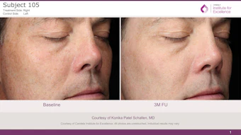
Patients treated with ablative fractional laser devices may experience dermal fibrosis following treatment while patients treated with nonablative devices do not, say researchers at the Netherlands Institute for Pigment Disorders, led by Bas A. Wind, MD, PhD. The study, which appeared in the journal of the American Society of Dermatologic Surgery (November 2011) involved 18 patients with pigment disorders. For each patient, two similar test regions were randomized for fractional laser treatment with intermittent topical bleaching and topical bleaching alone. Patients were further separated for treatment with either ablative or nonablative fractional lasers. Subjects with ashy dermatosis and post-inflammatory hyperpigmentation were treated with a 1550nm fractional device (15mJ/microbeam, 14% to 20% coverage); patients with Becker’s nevus were treated with an ablative CO2 fractional laser (10mJ/microbeam, 35% to 45% coverage). Both groups received three to five sessions, and bioposies were obtained three months after the last laser treatment. The researchers observed dermal fibrosis in four of eight patients treated using the ablative device and no patient who underwent the nonablative procedures. “Assuming that the dermal response is comparable in AD, PIH and BN, at the given settings, ablative FLT may induce fibrosis, whereas treatment with nonablative FLT does not. Whether formation of fibrosis has to be regarded as dermal remodeling or a subtle subclinical form of scarring should be investigated in future research,” concluded the study’s authors.
Fibrosis Found Following Fractional Laser Treatment
Feb 24th, 2012