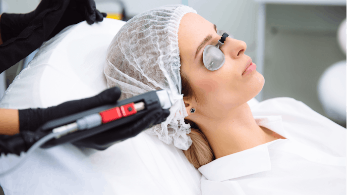
Research on the efficacy of medical aesthetic treatments relies heavily on patient-reported outcomes and feedback from blinded reviewers, but these measurements are subjective and may be unreliable. To counter these concerns, researchers Mikaela Kislevitz, BSN, RN, Karen B. Lu, MD, and colleagues at the UT Southwestern department of plastic surgery, investigated the use of microbiopsies and noninvasive imaging devices as potential tools to provide more objective data with low patient risk.
For their study, published in Lasers in Surgery and Medicine (November 2020), they assessed the outcomes of 12 laser skin resurfacing patients using the VISIA Skin Analysis System, high‐resolution ultrasonography, optical coherence tomography, BTC 2000 and microbiopsies.
Each patient received a single 1,470/2,940 nm laser treatment for facial rejuvenation. Assessments were performed before treatment and seven days, three weeks and three months post‐treatment. The VISIA Skin Analysis System was used to measure wrinkles, textures, pores, ultraviolet (UV) spots, brown spots, red areas and porphyrins. Other noninvasive skin measurements—high‐resolution ultrasonography, optical coherence tomography, transepidermal water loss and BTC 2000—were used to measure epidermal/dermal thickness, blood flow, surface roughness, wrinkle depth, attenuation coefficient, elasticity, laxity and viscoelasticity. The researcher performed 0.33 mm-diameter microbiopsies (the equivalent of a 23‐gauge needle) for histology and to measure gene expression of tissue rejuvenation.
The results revealed significant improvement in UV spots, brown spots and pore size three weeks after treatment and continued improvement in UV spots and brown spots three months after treatment. The dermal attenuation coefficient decreased significantly at three weeks, while blood flow 0.5 to 0.7 mm below the skin surface increased significantly between five days and three weeks following treatment. Epidermal hyaluronic acid expression (assessed by immunostaining and expression of inflammatory genes) was elevated seven days post‐treatment compared with untreated tissue and skin measurements taken three months post‐treatment. There were no statistically significant changes in collagen or elastin‐related genes between groups at the studied parameters. The microbiopsies left no scarring.
The authors concluded that “noninvasive devices can be effectively used to provide objective measurements of skin structure, pigmentation, blood flow and elasticity to assess the efficacy of facial skin rejuvenation treatments. Furthermore, microbiopsies can objectively evaluate facial skin rejuvenation without scarring.”
Read the full paper here.


