Bio-imaging methods are as complex as the skin structure and functions. Dermatology professionals usually decide aesthetic treatments based on clinical and or aesthetic visualization of skin. However, with current technological advances in bio-imaging, certain instruments can be very well incorporated in the derma-age management practice to understand skin’s cellular structure at a deeper level, allowing one to better manage the outcome of various aesthetic procedures. Also, skin bio-imaging offers clues on conditions that are beyond the aesthetic realm and may mandate medical intervention before starting aesthetic procedures. In this article, we will review eight instruments that can be helpful in planning aesthetic treatments.
1. Digital Photographic Imaging
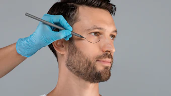
The simple principle behind the digital camera is to measure light reflection, refraction and scattering from the skin, and certain inferences are derived based on penetration and density of light through different skin tissues.
Polarizing filters that are attached to the camera’s lens separate reflectance and back scattering of light. A computer then breaks this electronic information down into digital data. Such parallel-polarized photography visualizes skin texture and scale. When this technique is used as cross-polarized photography, it enhances imaging of skin vascularity and pigmentation. Thus, one can determine the status and depth of skin pigmentation, and it can help in managing and planning age related pigmentation issues and treatments.
2. Dermatoscopy
Dermatoscopy or epiluminescence microscopy is the in vivo evaluation of epidermal microstructures, rete pegs, dermal papillae and papillary dermis. This device can assess skin structures in depth, and one can record skin images for before and after comparison and/or evaluation out of aesthetic procedures.
3. Confocal Microscopy
Confocal microscopy can provide better cellular resolution of skin and cutaneous structures. Confocal microscopes can create images by utilizing intrinsic differences between refractive indexes of cellular structures, especially melanin, collagen and keratin. Melanin and keratin provide contrast in confocal images. Melanin has the strongest refractive index of 1.7, and keratin’s refractive index is 1.5. Collagen and inflammatory cells are also other highly refractile structures. In the circumstances of targeting aesthetic revision of age-related wrinkles and instilling derma-fillers etc., this investigative method can be helpful in pointing potential skin areas to target.
4. Optical Coherence Tomography Optical Coherence (OCT)
Continue Reading our Digital Magazine to learn more about skin bio-imaging...Jayant Lokhande, M.D. BDP, is an Indian system of medicine practitioner and the chief scientific officer for Indus Extracts. He has formulated more than 50 dietary supplements, OTC drugs and functional foods and beverages worldwide. Lokhande earned both a M.D. BDP (botanical drug product) and a MBA in biotechnology. He has published two books, Handbook of Metallonutraceuticals and Botanical Drug Products: Recent Developments and Market Trends