Medtech company Enspectra Health published clinical trial results in JAMA Dermatology, assessing VIO, a handheld cross-modal imaging system, and its ability to visualize histologic skin features non-invasively, a process that typically requires invasive biopsy and histopathology. The study suggests that in addition to dermatology, the system's capabilities could serve personalized aesthetic assessment and monitoring.
This article is only available to registered users.
Log In to View the Full Article
Medtech company Enspectra Health published clinical trial results in JAMA Dermatology, assessing VIO, a handheld cross-modal imaging system, and its ability to visualize histologic skin features non-invasively, a process that typically requires invasive biopsy and histopathology. The study suggests that in addition to dermatology, the system's capabilities could serve personalized aesthetic assessment and monitoring.
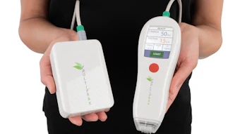
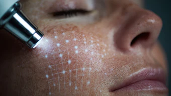
The VIO device uses reflectance confocal microscopy and multiphoton microscopy to generate cross-sectional, histology-like images of the skin. The 65 participants in the study were imaged with the cross-modal system [1]. Trained in dermatopathology, the physicians who used it achieved 96.4% accuracy in identifying primary histologic features and 98.5% accuracy for secondary features, per a Nov. 6 press release.
The study evaluated adults aged 18 to 99 years in two outpatient dermatology settings who were set to undergo lesional skin biopsy. VIO is FDA–cleared in assisting clinical judgment, leading to its implication in this VISTA Trial to create images of various tissue structures, including vessels, collagen, pigment and epidermal feature.
Focused on lesional skin, VIO's ability to visualize structural and cellular features non-invasively, the study states, has broad implications for aesthetics [1]. Cross-modal imaging may support pre-treatment assessment, post-treatment monitoring and personalized aesthetic planning by capturing changes in cellular architecture, collagen structure elastotic changes and pigmentation.
"During the trial, we imaged a range of common skin lesions across multiple anatomic locations and Fitzpatrick skin types I–V," said Michael Wang, MD, principal investigator for the study. "This design allowed us to evaluate the VIO system's ability to capture histologic features in a variety of commonly encountered clinical contexts."
While the system is not intended to diagnose disease or replace biopsy, and rather to assist with dermatology in clinical decision-making, VIO performs optical cross-sectional scanning to a depth of approximately 0.3 mm into the superficial dermis. The device emits low-power, single-wavelength laser pulses into the skin [1].
"These features are key to understanding skin health, and non-invasive visualization opens new possibilities for both medical and aesthetic applications," said Sarah Arron, MD, lead author of the study.
References:
1-https://jamanetwork.com/journals/jamadermatology/fullarticle/2841136