Osteopore International, a Singapore-maker of regenerative bone implants and Dr. Michael Wagels, deputy director of plastic and reconstructive surgery at Princess Alexandra Hospital in Brisbane, Australia, have announced the successful outcome of a first-of-its-kind cranioplasty procedure using a 3D printed bioresorbable polycaprolactone (PCL) regenerative bone implant.
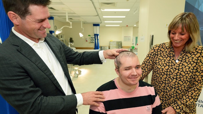
The patient, 26-year-old Brodie Ellis, suffered a Stage 4 brain injury and a severely broken leg from a motorcycle accident in Vietnam in December 2018. On top of having to amputate his left leg, certain parts of his skull had to be removed and replaced with plastic implants. Unfortunately, according to Dr. Wagels, "one of the [plastic] implants [had become] exposed and developed an infection. Because the implant had no blood supply, the infection just kept getting worse and worse, so it had to be removed. This left Brodie with headaches and a contour deformity of the skull."
To treat these problems, Dr. Wagels recommended replacing a section of missing skull with Osteopore's 3D printed bioresorbable polycaprolactone (PCL) regenerative bone implant. This PCL bone implant would have the ability to encourage natural bone regrowth before disintegrating into carbon dioxide and water with no foreign material left in the skull, thereby reducing post-surgical or removal surgery complications and significantly minimizing the risk of infection.
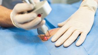
The 11-hour surgery involved implantation using an innovative surgical technique that transplants a tissue flap from the patient's knee and required a customized implant, which was designed by the Osteopore team using the patient's CT scan in consultation with Dr. Wagels. Sitting perfectly on the contour of Ellis' skull, the biomimetic structure of the 3D printed regenerative implant introduced a patented, interconnected porous scaffold that mimics the natural cancellous bone microstructure. This mesh-like structure promotes blood vessel infiltration to support bone and tissue regrowth. The PCL material of the implant is designed to then be gradually resorbed and metabolised by the body over an 18 to 24 months period.
Just weeks after the cranioplasty surgery, CT scans of Ellis' skull showed that the implant had successfully enabled new bone to form within the scaffold. “The latest CT scan taken eight weeks after the operation shows bone forming both on the outside and inside of the implant, indicating the body has recognized the implant as broken bone that needs to be healed,” said Dr. Wagels.
Less than a month after the cranioplasty, Ellis was discharged from Princess Alexandra Hospital, and has been able to function independently at home while regaining his strength at the gym.