If you work with pigmented skin, you have probably seen this happen. A client comes in with a facial patch, you assume you know what it is, you prescribe the protocol that usually works, and three weeks later she is back with the spot darker than when she started. She is frustrated, and she is starting to doubt you.
Melasma and solar melanosis (solar lentigines) can look alike enough to fool you, especially when you are moving fast. Biologically, though, they are very different processes, and getting the diagnosis wrong does more than waste time. It can make the pigmentation worse.
The Two Spots, Side by Side
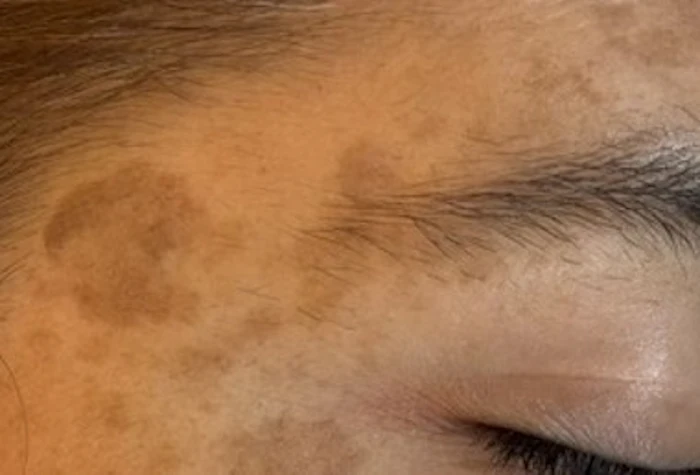
The images below show the core visual difference. The clue I trust most on a first exam is the distribution.
 Airton Barbalho Rodrigues
 Melasma: confluent, symmetric patches with poorly defined borders, concentrated in the malar region. This is the same patient as Figure 1, a mixed presentation that shows how often the two conditions coexist in everyday practice.
Two Different Biologies
Melasma is a matter of melanocyte hyperfunction. The melanocytes do not necessarily multiply. They become hyperactivated and produce melanin in a dysregulated way. The usual triggers are UV radiation, hormones (pregnancy, oral contraceptives) and visible light. What matters most clinically is that inflammation can drive further pigment production. An aggressive laser, a strong peel or other trauma may induce post-inflammatory hyperpigmentation in susceptible patients, so the intervention meant to clear the pigment can end up deepening it. [1,2]
Solar melanosis is a matter of melanocyte hyperplasia from cumulative sun damage. Chronic UV exposure drives melanocytes to multiply and activate. The lesions are discrete and well-demarcated, without facial symmetry and without a hormonal component, and these cells do not carry the same tendency to worsen with controlled inflammation. [4]
That microscopic difference is what separates a good outcome from a bad one once you are in the treatment room.
How to Tell Them Apart in Practice
Distribution pattern, a simple clinical observation:
• Melasma: symmetric, bilateral, concentrated in the malar, centrofacial or mandibular area, with poorly defined borders and confluent patches.
• Solar melanosis: isolated, discrete, well-demarcated lesions, randomly distributed in chronically photo-exposed areas, with no symmetry.
Clinical history, always ask:
• Melasma: a history of pregnancy, oral contraceptive use or hormone therapy, and worsening with sun exposure.
• Solar melanosis: prolonged occupational or recreational sun exposure, with generalized photoaging.
Wood's lamp, and what is backing it up today
The classic teaching is that a Wood's lamp examination separates epidermal from dermal pigment. Melasma with a strong epidermal component looks markedly darker under the light, while solar melanosis tends to show a flatter, more uniform pattern. It is still a fast bedside check worth doing, since it costs nothing and takes about thirty seconds.
It is worth being honest about its limits, though. Recent literature has found Wood's lamp results to be inconsistent, especially in darker skin phototypes, which is exactly where melasma is most common. Current guidance no longer treats it as a reliable basis for prognosis or treatment on its own. [1] Dermoscopy has become a useful next step in trained hands, helping read pigment network and vascular patterns that the naked eye misses.
This is also why digital skin analyzers have become standard equipment in many practices. Polarized and UV-filtered imaging gives a more standardized, reproducible view of pigment distribution than a Wood's lamp alone, and it leaves you with a documented image you can compare over time. More recently, AI-assisted image analysis has entered the picture, with software that flags pigment patterns and proposes a differential. In my experience these tools are a useful second opinion, particularly for a newer practitioner facing an atypical or mixed case. They come with a real caveat, though: across dermatology more broadly, AI accuracy has been shown to drop on darker skin tones, largely because the datasets used to train these models underrepresent skin of color. [5] I read an analyzer or an AI suggestion the same way I read the Wood's lamp, as one more piece of information rather than the verdict.
When Both Appear Together
The patient in Figures 1 and 2 is a good reminder that mixed pigmentation is more common than isolated disease. Years of sun exposure and a hormonal trigger often land on the same face, and you end up looking at confluent malar melasma alongside a scatter of discrete lentigines. Treat that face as if it were pure lentigo and you risk inflaming the melasma; treat it as if it were pure melasma and the lentigines never fully clear.
The practical move is to separate the two in your mind before you plan anything. Map which areas are confluent and symmetric (melasma) and which are discrete and well-demarcated (lentigines). Then sequence the work: stabilize and treat the melasma first with photoprotection and topicals, get it quiet, and only then address the lentigines with a more direct approach. Combining protocols is possible, but it belongs later, once the melasma is under control and you know the skin is not reacting with rebound pigment.
The Cost of Getting It Wrong
The same protocol applied to the wrong diagnosis produces two opposite outcomes, and both are costly:
Error 1. High Risk: Melasma Treated as Solar Melanosis
- Stimulus: high-fluence laser or aggressive peel
- Reaction: thermal inflammation and tissue trauma
- Cellular response: further melanin production in susceptible skin
- Outcome: severe post-inflammatory hyperpigmentation
Error 2. By Omission: Solar Melanosis Treated With Excessive Caution
- Approach: mild topicals only, hesitant to intervene
- Reality: the lesion would have tolerated a definitive protocol
- Consequence: treatment drags on for months
- Outcome: the chance at a clean, lasting result is lost
What separates those two outcomes is not the technology. It is the diagnosis you make before you switch the device on.
Diagnosis-Based Protocol
What follows reflects both the current literature and my own clinical protocol, organized by therapeutic line. [1,3]
If Melasma:
• First line: rigorous photoprotection (SPF 50+, reapplied every two hours, tinted or physical blocker) plus topical depigmenting agents (hydroquinone, vitamin C, azelaic acid).
• Second line: gentle, superficial chemical peels, added cautiously once the skin is stable.
• Third line: low-aggressiveness laser, reserved for refractory cases and only after photoprotection and topicals are optimized.
If Solar Melanosis:
• First line: standard photoprotection.
• Second line: moderate-to-strong chemical peels.
• Third line: Q-switched laser, with the expectation of a faster, more definitive result and lower recurrence risk.
Five Questions Before You Treat
Before reaching for any device, I run through the same five questions. They take under two minutes and settle most cases:
1. Is the pigmentation symmetrical?
2. Is there a hormonal history (pregnancy, contraceptives, hormone therapy)?
3. Are the borders well-defined, or diffuse?
4. Is the involvement focal and discrete, or confluent?
5. Has a previous treatment made the pigmentation worse?
Diffuse, symmetric, hormone-linked pigmentation that has flared after treatment points to melasma. Discrete, well-defined, sun-driven lesions with no such history point to solar lentigines. When the answers pull in both directions, you are almost certainly looking at a mixed case.
The Details That Make the Difference
The Wood's lamp takes thirty seconds. The clinical history takes two minutes, and reading the distribution pattern takes one more. Less than five minutes stand between a confident treatment plan and a protocol that could make everything worse.
The responsibility here is not only technical. It is clinical. Clients often arrive already frustrated because someone got it wrong before, and that erodes their trust in the whole profession. In most of those cases, careful differential diagnosis based on simple observation would have been enough to avoid the problem.
Successful pigment treatment begins long before the laser is switched on. The most important instrument in the treatment room is still the clinician's ability to recognize the biology behind the discoloration.
References
1. Sathe NC, Launico MV. Melasma. StatPearls [Internet]. Treasure Island (FL): StatPearls Publishing; 2026 Jan. Bookshelf ID: NBK459271. PMID: 29083744.
2. Sheth VM, Pandya AG. Melasma: a comprehensive update: part I. J Am Acad Dermatol. 2011;65(4):689-697.
3. Sheth VM, Pandya AG. Melasma: a comprehensive update: part II. J Am Acad Dermatol. 2011;65(4):699-714.
4. Lee WJ, Jo SY, Lee MH, et al. The Effect of MCP-1/CCR2 on the Proliferation and Senescence of Epidermal Constituent Cells in Solar Lentigo. Int J Mol Sci. 2016;17(6):948.
5. Liu Y, Primiero CA, Kulkarni V, Soyer HP, Betz-Stablein B. Artificial Intelligence for the Classification of Pigmented Skin Lesions in Populations with Skin of Color: A Systematic Review. Dermatology. 2023;239(4):499-513.