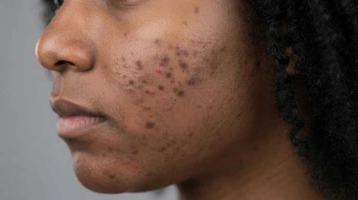
Post-inflammatory hyperpigmentation (PIH) is often misunderstood as a simple pigment problem. In reality, it is the visible consequence of a broader response to injury. For many patients, the wound heals within weeks, yet the discoloration that follows can persist for months or even years and may become more emotionally distressing than the original trauma. This is especially relevant in darker skin phototypes, where inflammatory stimuli can produce a stronger and more persistent pigmentary response [1,2].
In practice, patients come worried because the mark remains visible and because they do not know whether it will fade. This is why PIH deserves a more complete clinical approach than simply waiting for the color to improve.
Related: Oral Tranexamic Acid Helps Prevent PIH
The first days after injury matter because the future behavior of pigmentation may be influenced before the dark mark is fully visible. During early repair, the skin is not only closing a wound; it is coordinating inflammation, vascular signaling, oxidative stress, keratinocyte-melanocyte communication, extracellular matrix repair, and restoration of the dermoepidermal junction. If this microenvironment remains inflamed, oxidatively stressed, or poorly protected from ultraviolet and visible light, melanocyte stimulation may persist beyond the initial healing process and contribute to more resistant discoloration [1,9].
The typical progression begins with tissue damage and inflammation. Inflammatory mediators, reactive oxygen species, vascular signals, and growth factors can stimulate melanocytes to increase melanin production. This response is protective by nature, but in susceptible individuals it may become excessive. When pigment remains mainly epidermal, improvement is often faster. When inflammation disrupts the epidermal-dermal interface and pigment drops into the dermis, commonly described as pigment incontinence, the discoloration may appear deeper, grayer, and slower to clear [1,3].
Related: Bakuchiol Reduces Acne Blemishes and PIH
Timing is one of the most important clinical decisions. Historically, many clinicians adopted a “wait and see” approach until the scar had fully matured. Today, the approach is more nuanced. Aggressive pigment treatments should be avoided during active wound healing, but early supportive management after safe re-epithelialization is rational. The goal is not to treat the pigment aggressively while the tissue is still repairing; it is to reduce the inflammatory, oxidative, vascular, and photoinduced signals that may drive pigment persistence [1,4].
For this reason, the first phase of management should begin with barrier restoration, inflammation control, oxidative stress reduction, photoprotection, and low-irritation pigment regulation, not exfoliation or aggressive pigment suppression.
It is important to select ingredients whose concentration, vehicle, and mechanism of action are compatible with healing skin and do not increase barrier disruption or irritation. In this setting, antioxidant and melanoregulatory ingredients such as ferulic acid, grape seed extract, thioctic acid, licorice extract, niacinamide, tranexamic acid, and phytic acid may be useful because they support oxidative balance, calm inflammatory signaling, and help regulate pigment without relying primarily on irritation or forced exfoliation. Gentle cleansing is essential; non-stripping cleansers and mild surfactant systems are preferable, while scrubs, exfoliating acids, harsh soaps, fragrance-heavy products, and unnecessary irritants should be avoided. Furthermore, when clinically indicated, short-term physician-directed anti-inflammatory therapy such as topical corticosteroids or other non-irritating anti-inflammatory approaches may be considered. Adjunctive modalities such as LED light therapy may also be useful in selected cases to support inflammation control and recovery. All these options should be individualized according to wound status, anatomical site, skin phototype, and risk of irritation. The key is not only the ingredient itself, but its concentration, vehicle, timing, and the patient’s tolerance. [1,5].
Photoprotection remains the most evidence-supported intervention throughout the entire treatment journey. Ultraviolet radiation and visible light can prolong pigmentation by continuing to stimulate melanocyte activity. Broad-spectrum sunscreen is therefore not an accessory; it is part of treatment. In patients prone to hyperpigmentation, especially darker phototypes, tinted sunscreens containing iron oxides may provide additional protection against visible light. Physical measures, like shade, hats, protective clothing, and covering the affected area when possible are equally important because sunscreen alone cannot compensate for repeated high- intensity exposure during the vulnerable repair phase [4,6,7,8].
Once the barrier is stable, treatment can progress toward pigment modulation and epidermal renewal. Although many clinical studies evaluate therapies as monotherapy, PIH is not a single-pathway condition; therefore, a logical clinical approach often involves synergistic combinations that address melanogenesis, inflammation, oxidative stress, epidermal turnover, barrier function, and, in some cases, dermal pigment deposition. Renewal-oriented actives such as mandelic acid, salicylic acid, retinoids, and other acids may have a role at this stage, especially when combined with more targeted pigment-regulating agents such as arbutin, or with previously introduced melanoregulatory ingredients such as tranexamic acid, azelaic acid, vitamin C derivatives, or niacinamide that may now be used at higher concentrations, greater frequency, or in more corrective protocols according to skin tolerance. These agents should be selected according to skin type, phototype, sensitivity, pigment depth, and tolerance, and introduced progressively to avoid secondary irritant dermatitis that could worsen the clinical outcome. The key is not simply choosing ingredients that “lighten pigment,” but selecting the right agents,vehicles, concentrations, frequencies, and sequences to correct discoloration without creating new inflammation [1,2].
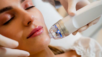
For more persistent discoloration, in-office procedures may be introduced progressively. Chemical peels, microneedling, targeted transdermal delivery systems, and selected laser-based technologies can accelerate improvement when appropriately indicated. However, evidence requires caution: in skin of color, partial improvement is more common than complete clearance, and overly aggressive
parameters can worsen pigmentation. Procedures should therefore be introduced as part of a guided sequence, not as isolated interventions. A peel, microneedling session, or laser treatment performed on inflamed, barrier-impaired, or photosensitized skin may amplify the very pathway it is intended to correct [2,5,12].
The exposome also matters. Ultraviolet exposure, visible light, pollution, heat, sleep deprivation, smoking, nutrition, psychosocial stress, and repeated irritation can contribute to oxidative stress and inflammatory signaling in the skin. While not all exposome-related factors have been studied specifically in post-traumatic PIH, they are relevant to pigmentation, inflammation, barrier health, and skin aging. For patients, this means that success depends not only on what is applied in the office or at home, but also on how the skin is protected in daily life [10,11].
A critical aspect of management is determining risk rather than deciding whether the case “deserves” treatment. Risk assessment should not make the clinician passive or overly conservative. On the contrary, it helps define prognosis and guide an effective preventive strategy. Because hyperpigmentation can significantly affect self-perception and quality of life, prevention and early supportive management should be considered legitimate care, not cosmetic overreaction [1,2,13].
Equally important is setting realistic expectations. PIH improves gradually because the skin must clear accumulated pigment while restoring balance. Epidermal pigmentation may improve over weeks to months, while deeper dermal pigment can take longer and may be more resistant. Most patients require several months of consistent therapy, and resistant cases may need long-term maintenance. Premature escalation can increase irritation and delay recovery [1,2].
The most successful outcomes occur when clinicians recognize that persistent discoloration is not merely a pigment disorder, but part of a broader healing process involving inflammation, barrier function, vascular signaling, scar remodeling, oxidative balance, extracellular matrix integrity, and cellular communication. This is the philosophy that guides my approach within the SURPLEXION ecosystem: preparation, recovery, correction, and maintenance.
This is where systems such as Complexion Code and INFUSION SQ become clinically useful. They allow the clinician to adapt intensity, timing, and delivery according to the skin’s phase of recovery. Complexion Code supports the topical side of the journey with strategies that can be adapted across phases: antioxidant defense, barrier comfort, tone regulation, progressive pigment correction, and long-term maintenance through carefully selected combinations of melanoregulatory, renewing, and restorative ingredients. Its different options within the range make it possible to adjust the level of intervention according to skin tolerance, timing, phototype, and inflammatory status, which is essential when treating PIH-prone skin.
INFUSION SQ extends this approach into the in-office setting through precision delivery and targeted skin quality protocols. Rather than treating pigment as an isolated endpoint, it supports the broader tissue environment that influences visible quality, including tone, texture, firmness, radiance, oxidative balance, and tissue-level communication. Together, Complexion Code and INFUSION SQ reflect the same clinical principle: effective PIH management is not defined by one aggressive intervention, but by the right sequence, the right target, and the right level of stimulation for the skin’s current state.
This systems-based approach requires clinical judgment. The clinician must decide when the skin is ready, which pathways need priority, and how much stimulation the tissue can safely tolerate. Early management should focus on protection and recovery; later phases may progress toward pigment modulation, texture improvement, scar quality, and maintenance. Ultimately, modern PIH care is moving beyond simply treating a dark spot and toward guiding the skin through a complete recovery process one based on earlier recognition, better sequencing, strong photoprotection, low-irritation correction, and respect for the biology of healing skin.
Practical step-by-step guide from clinical experience
1. First, assess risk not to become overly conservative, but to define prognosis, communicate realistic expectations, and guide an effective
preventive strategy. Evaluate phototype, intensity of inflammation, location, depth and color of pigment, scar behavior, sun exposure, history of irritation, and patient distress.
2. Do not treat aggressively over an open wound or actively inflamed skin. Wait until safe re-epithelialization before introducing active pigment care.
3. In the early phase, prioritize gentle cleansing, barrier repair, anti-inflammatory support, antioxidant protection, strict photoprotection, and low-irritation melanoregulatory ingredients that are compatible with healing skin.
4. Avoid friction, heat exposure, early exfoliation, harsh acids, irritating vehicles, aggressive retinoids, and unnecessary procedures while the skin remains reactive.
5. Once the barrier is stable, intensify pigment modulation progressively by combining melanoregulatory agents with epidermal renewal strategies, adjusting concentration, frequency, and vehicle according to tolerance.
6. In persistent PIH, consider in-office procedures only after adequate preparation: superficial peels, microneedling/TDS, or carefully selected lasers depending on phototype, pigment depth, scar quality, vascular/inflammatory activity, and risk of rebound pigmentation.
7. Maintain results with daily photoprotection, barrier support, antioxidant care, gentle pigment regulation, and periodic reassessment. In PIH, recurrence prevention is part of treatment.
For more information on Complexion Code, INFUSION SQ, or other systems referenced in this article, visit xtetic.com.
References
1. Davis, E. C., & Callender, V. D. (2010). Postinflammatory hyperpigmentation: A review of the epidemiology, clinical features, and treatment options in skin of color. The Journal of Clinical and Aesthetic Dermatology, 3(7), 20 31.
2.Mar, K., Khalid, B., Maazi, M., Ahmed, R., Wang, O. J. E., & Khosravi-Hafshejani, T. (2024). Treatment of post-inflammatory hyperpigmentation in skin of colour: A systematic review. Journal of Cutaneous Medicine and Surgery, 28(5), 473 480. https://doi.org/10.1177/12034754241265716
3.DermNet. (n.d.). Postinflammatory hyperpigmentation. DermNet NZ. https://dermnetnz.org/topics/postinflammatory-hyperpigmentation
4. Mar, K., Maazi, M., Khalid, B., Ahmed, R., Wang, O. J. E., & Khosravi- Hafshejani, T. (2025). Prevention of post-inflammatory hyperpigmentation in skin of colour: A systematic review. Australasian Journal of Dermatology, 66(3), 119
126. https://doi.org/10.1111/ajd.14432
5. Amechi, M., & Halpin, J. (2023). Considerations for laser therapy, microneedling, and chemical peels when treating patients with skin of color. Plastic and Aesthetic Nursing, 43(1), 14 21. https://doi.org/10.1097/PSN.0000000000000483
6. Fatima, S., Braunberger, T., Mohammad, T. F., Kohli, I., & Hamzavi, I. H. (2020). The role of sunscreen in melasma and postinflammatory
hyperpigmentation. Indian Journal of Dermatology, 65(1), 5 10. https://doi.org/10.4103/ijd.IJD_295_18
7. Lyons, A. B., Trullas, C., Kohli, I., Hamzavi, I. H., & Lim, H. W. (2021). Photoprotection beyond ultraviolet radiation: A review of tinted sunscreens. Journal of the American Academy of Dermatology, 84(5), 1393 1397. https://doi.org/10.1016/j.jaad.2020.04.079
8. Tsai, J., Chien, A. L., & Lim, H. W. (2022). Photoprotection for skin of color. American Journal of Clinical Dermatology, 23(2), 195 205.
https://doi.org/10.1007/s40257-021-00670-z
9.Markiewicz-Tomczyk, A., Budzisz, E., & Erkiert-Polguj, A. (2023). A subjective and objective assessment of combined methods of applying chemical peels and microneedling in antiaging treatments. Journal of Clinical Medicine, 12(5), 1869. https://doi.org/10.3390/jcm12051869
10. Papaccio, F., D’Arino, A., Caputo, S., & Bellei, B. (2022). Focus on the contribution of oxidative stress in skin aging. Antioxidants, 11(6), 1121. https://doi.org/10.3390/antiox11061121
11. Krutmann, J., Bouloc, A., Sore, G., Bernard, B. A., & Passeron, T. (2017). The skin aging exposome. Journal of Dermatological Science, 85(3), 152 161. https://doi.org/10.1016/j.jdermsci.2016.09.015
12. Sowash, M., & Alster, T. S. (2023). Review of laser treatments for post- inflammatory hyperpigmentation in skin of color. American Journal of Clinical Dermatology, 24(3), 381 396. https://doi.org/10.1007/s40257-023-00759-7